
Advanced Gastroenterology Care - Every part of your GUT
Comprehensive digestive care with compassion and cutting-edge technology






ABOUT US
Welcome to Shri Siddhivinayak Polyclinic
- Seamless Care
- Patient-Centered Care
- Comprehensive Care
- Pain Free
- Scarless Endoscopic Solutions
- 24/7 Support
Meet Our Experts
Gastroenterology & Advanced Endoscopy
Dr. Gaurav Patil
Director
Pancreatobiliary Clinic
Key Conditions Treated:
Read MoreLess
Pancreatitis is the inflammation of the pancreas, an organ that produces digestive juices to help break down food in the small intestine and insulin to regulate blood sugar levels.
Healthy vs. Affected Pancreas
In acute pancreatitis, the pancreas becomes inflamed and may show signs of hemorrhaging compared to a healthy pancreas.
Symptoms of Acute Pancreatitis
The primary symptom is abdominal pain, ranging from mild to severe. Patients also typically have elevated pancreatic enzymes, detectable in blood and urine tests.
Other possible symptoms include:
- Nausea and vomiting
- Fever
- Mild jaundice
- Confusion
- Respiratory failure
Diagnosis of Acute Pancreatitis
Diagnosis begins with a physical examination and a detailed discussion of medical history and symptoms. Doctors may order several tests, including:
1. Laboratory Testing
- During an acute attack, the pancreas releases enzymes into the blood.
- Amylase and lipase levels are the most commonly measured.
- These are checked through blood and urine tests.
2. Imaging Studies
Imaging is recommended for patients with severe abdominal pain. These studies can reveal:
- Obstructions
- Fluid buildup
- Increased separation between the stomach and colon (a sign of inflammation)
Common imaging methods:
- Ultrasound: Uses sound waves to create images. Not very sensitive for detecting pancreatic changes, but helpful for identifying gallstones and obstructions.
- Computed Tomography (CT) Scan: A powerful X-ray and the most reliable diagnostic test for acute pancreatitis. A contrast-enhanced CT scan is often used to highlight abnormalities.
3. Endoscopic Procedures
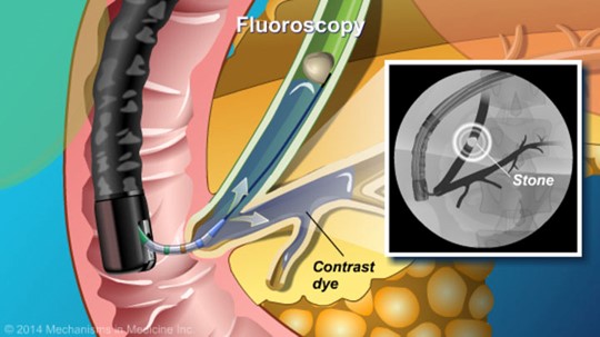
- Endoscopic Retrograde Cholangiopancreatography (ERCP): A specialized test that combines endoscopy and X-ray imaging.
- ERCP with Sphincter of Oddi Manometry: Measures pressure in the bile and pancreatic ducts, helping diagnose underlying causes of pancreatitis.
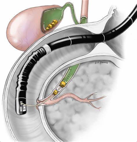
Gallstone pancreatitis happens when a gallstone blocks the tube (duct) that drains the pancreas. This causes the pancreas to become inflamed and painful. The pancreas makes insulin (controls blood sugar) and enzymes (helps digest food).
Causes
- Gallstones form in the gallbladder.
- If a stone moves and blocks the duct between the pancreas and small intestine, it can cause a backup of fluid → leading to inflammation and pain.
- Gallstones are the most common cause of sudden (acute) pancreatitis.
Symptoms
Common signs include:
- Severe belly pain (upper or left side, may spread to the back, chest, or shoulder)
- Nausea or vomiting
- Fever and chills
- Yellowing of skin or eyes (jaundice)
- Pain that feels sharp, squeezing, or cramping
Diagnosis
- Blood tests → to check for inflammation.
- Imaging tests (CT scan, MRI, or ultrasound) → to see if a stone is stuck in the duct.
Treatment
Most people need hospital care. Treatment may include:
- IV fluids to keep you hydrated.
- IV medicines for pain and nausea.
- Special feeding support if you can’t eat.
If the stone does not pass on its own:
- ERCP (a scope procedure to remove the stone).
- Surgery to remove the stone or gallbladder.
⚠️ Gallbladder removal (cholecystectomy) is often recommended to prevent the problem from happening again.
Complications (If Untreated)
- Infection (cholangitis)
- Jaundice
- Death of pancreas tissue (pancreatic necrosis)
- Can be life-threatening
Prevention
- You can lower your risk of gallstones by:
- Eating a healthy diet
- Staying at a healthy weight
- Managing cholesterol and diabetes
- Talking to your doctor about medications (like estrogen) that may increase risk
Once you’ve had gallstone pancreatitis, gallbladder removal is the best way to prevent it from coming back.
When to Call Your Doctor
Get immediate medical help if you have:
- Severe belly pain
- Fever or chills
- Jaundice (yellow skin or eyes)
- Nausea and vomiting
What Are Gallstones?
- Gallstones form when bile (a digestive fluid stored in the gallbladder) hardens into stone-like material.
- They may be made of cholesterol, bile salts, or bilirubin (a pigment from red blood cells).
Types:
- Cholelithiasis: Gallstones in the gallbladder.
- Choledocholithiasis: Gallstones in the bile ducts.
⚠️ Gallstones that block bile ducts can cause serious or life-threatening infections of the liver, pancreas, or bile ducts.
Causes
- Cholesterol stones: Too much cholesterol or bilirubin, not enough bile salts, or poor gallbladder emptying.
- Pigment stones: More common in people with cirrhosis, biliary infections, or blood disorders (like sickle cell anemia).
Symptoms
Many gallstones cause no symptoms (“silent stones”).
When symptoms occur, they may include:
- Sudden, severe pain in the upper abdomen (often after fatty meals or at night)
- Pain between the shoulder blades or in the right shoulder
- Nausea or vomiting
- Fever and chills
- Jaundice (yellowing of skin/eyes)
- Bloating, gas, belching, or indigestion
Seek immediate medical help if you have:
- Sweating and chills
- Low-grade fever
- Yellow skin or eyes
- Clay-colored stools
Who Is at Risk?
You may be more likely to develop gallstones if you have:
- Obesity (especially women)
- High estrogen levels (pregnancy, hormone therapy, birth control pills)
- Native American ancestry (higher genetic risk)
- Female gender (twice the risk of men)
- Age over 60
- Cholesterol-lowering drugs (increase cholesterol in bile)
- Diabetes (high triglycerides)
- Rapid weight loss or fasting (causes bile to become concentrated)
Diagnosis
Gallstones are sometimes found by accident during other tests. If you have symptoms, your doctor may order:
- Ultrasound (most common test)
- X-ray (cholecystography) with contrast dye
- Blood tests (check for infection, jaundice, or pancreatitis)
- CT scan (detailed cross-section images)
- ERCP (scope test with dye to see bile ducts and sometimes remove stones)
- Sphincterotomy (widening the bile duct opening to let stones pass)
Treatment
If gallstones cause no symptoms, treatment may not be needed.
If they do cause problems, options include:
Gallbladder removal (cholecystectomy)
- The most common treatment.
- Bile will flow directly from the liver to the intestine.
- Some people may have mild diarrhea afterward.
Medicines to dissolve stones
- Oral bile acid drugs (slow process, not always effective).
Special procedures
- Methyl-tert-butyl ether injection (dissolves stones).
- Contact dissolution therapy (experimental, injects drugs directly into gallbladder).
What Are Bile Duct Stones?
Bile duct stones are hardened deposits that form in the bile ducts, which are tubes carrying bile from the liver and gallbladder to the small intestine.
Stones can block bile flow, causing pain, infection, or liver problems.
Causes of Bile Duct Stones
Stones that move from the gallbladder into the bile ducts
Sometimes stones form directly in the bile ducts (less common)
Risk factors include gallstones in the gallbladder, obesity, certain medications, and liver disease
Symptoms of Bile Duct Stones
You may experience:
Pain in the upper right abdomen
Jaundice (yellowing of skin or eyes)
Fever or chills
Nausea and vomiting
Dark urine or pale stools
Diagnosis
Doctors may use:
Ultrasound – to look for stones in the bile ducts
CT or MRI scans – to get detailed images
ERCP (Endoscopic Retrograde Cholangiopancreatography) – a procedure to see and sometimes remove stones
Blood tests – to check liver function and look for infection
Treatment
Endoscopic removal – stones can often be removed using a scope (ERCP)
Surgery – in some cases, surgery may be needed to remove stones
Medications – to dissolve stones in rare situations
What Are Bile Duct Tumors?
Bile duct tumors are abnormal growths in the bile ducts. They may be benign (non-cancerous) or malignant (cancerous).
Causes and Risk Factors
Chronic inflammation of the bile ducts
Bile duct cysts
Certain liver diseases
Age and family history may increase risk
Symptoms of Bile Duct Tumors
Jaundice (yellow skin or eyes)
Abdominal pain
Itching
Unexplained weight loss
Fever or night sweats (less common)
Pale stools or dark urine
Diagnosis
Imaging tests: Ultrasound, CT, or MRI
ERCP: Can be used to take a tissue sample (biopsy)
Blood tests: May show liver function changes or tumor markers
Treatment
Surgery – removal of the tumor if possible
Chemotherapy or radiation – may be recommended for malignant tumors
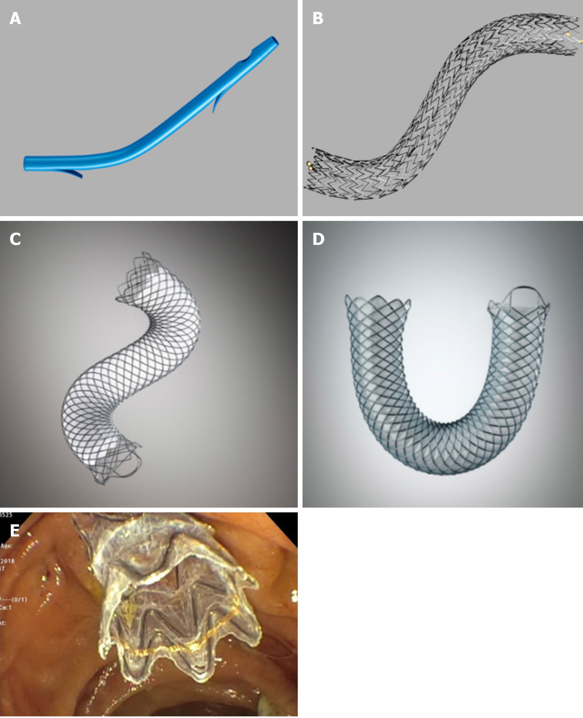
Stent placement – to keep the bile duct open if blocked
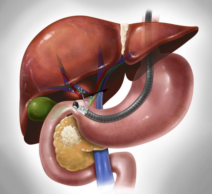
Advanced Procedures:
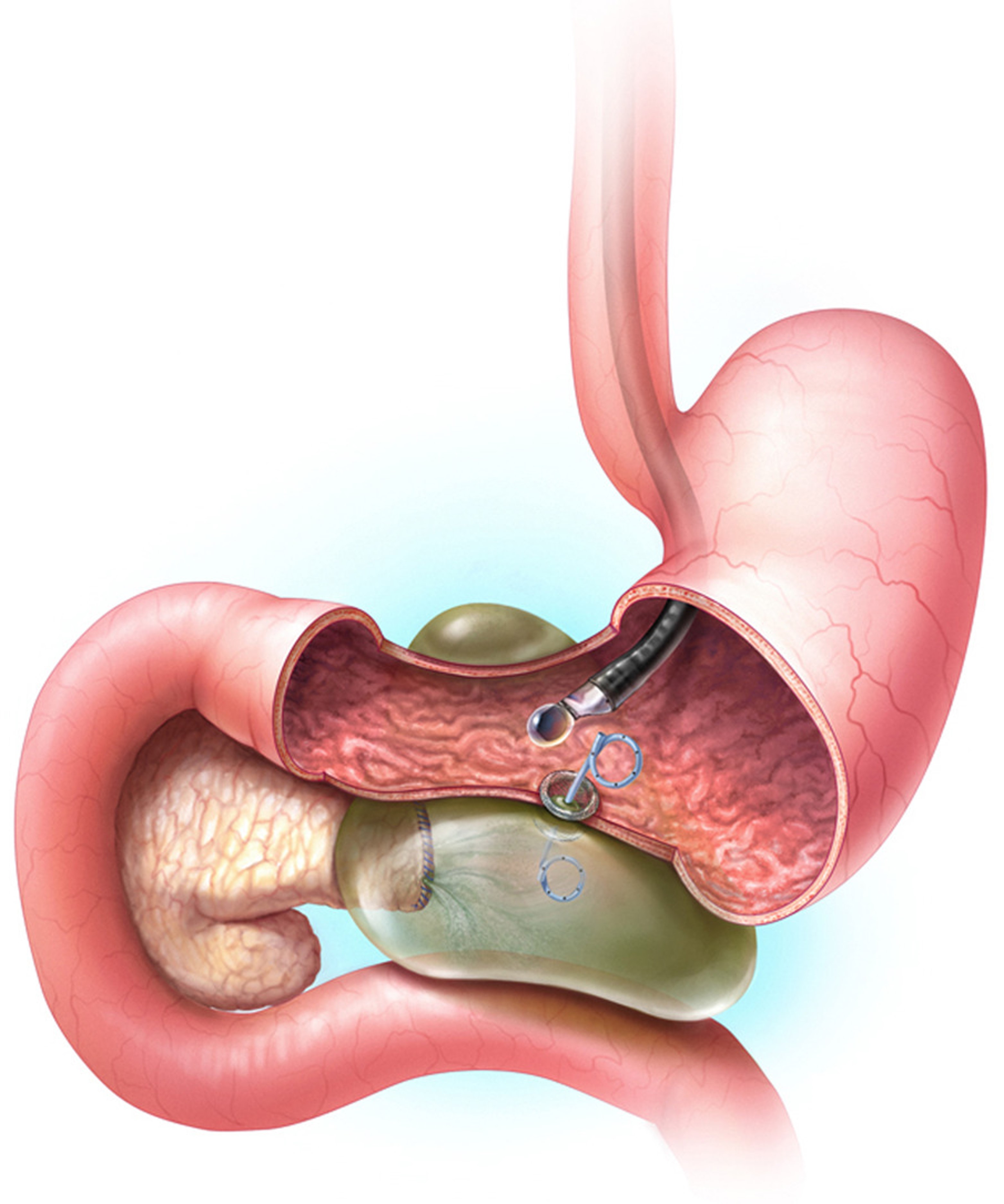
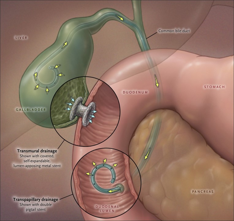
Flexible Endoscopic Surgery & Oncology Clinic
Key Conditions Treated:
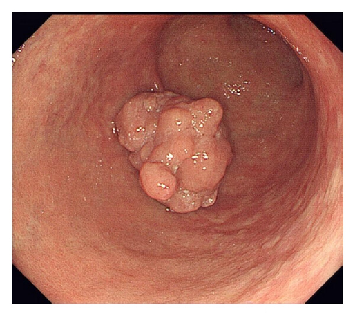
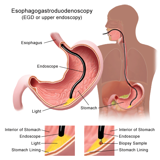
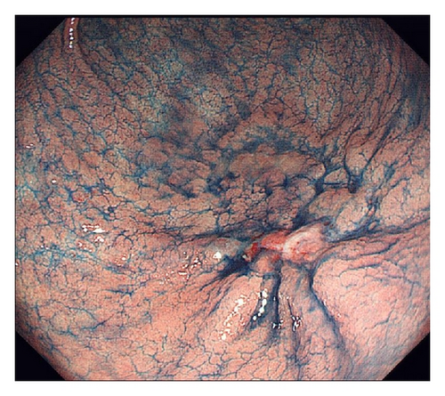
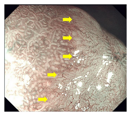
Stomach Cancer
Read MoreLess
What Is Stomach Cancer?
Stomach cancer (also called gastric cancer) begins in the stomach.
The most common type is adenocarcinoma, which starts in the stomach’s inner lining.
Other types exist, but they are less common.
While cases have declined in recent years, it remains a major cancer worldwide.
What Causes It?
The exact cause is not fully known, but certain risk factors increase the chances:
Diet: Eating lots of smoked, salted, or processed meats and too few vegetables.
Lifestyle: Smoking and drinking alcohol.
Helicobacter pylori infection: A bacteria that can damage the stomach lining.
Other stomach conditions: Sores (ulcers) or abnormal growth of stomach lining cells.
Family history: Certain inherited conditions.
⚠️ Stomach cancer can still occur even without these risk factors.
Symptoms
Early stomach cancer may cause no symptoms or only mild ones, such as:
Feeling full quickly (early satiety)
Mild stomach discomfort
Tiredness (fatigue)
More concerning symptoms include:
Unexplained weight loss
Severe belly pain
Nausea and vomiting
Loss of appetite
Vomiting blood
Dark, sticky stools (tarry stool)
Trouble or painful swallowing
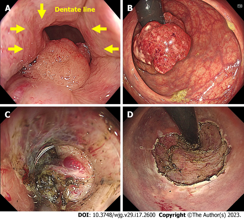
What Is Colorectal Cancer?
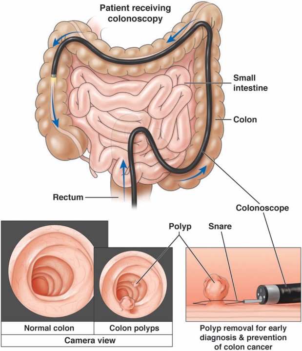
Colorectal cancer is cancer of the colon or rectum.
It is the second leading cause of cancer death in the United States.
About 56,000 people die from it each year.
Symptoms
Colorectal cancer often causes no symptoms in the early stages. Symptoms usually appear as the disease progresses.
Possible warning signs include:
Blood in the stool (sometimes not visible)
Anemia (low red blood cell count)
Abdominal discomfort or cramping
Nausea and vomiting
Pelvic pain
Unexplained weight loss
Changes in bowel habits
Unusual diarrhea or constipation
Thinner than normal stools
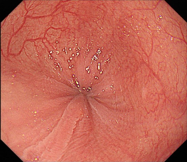
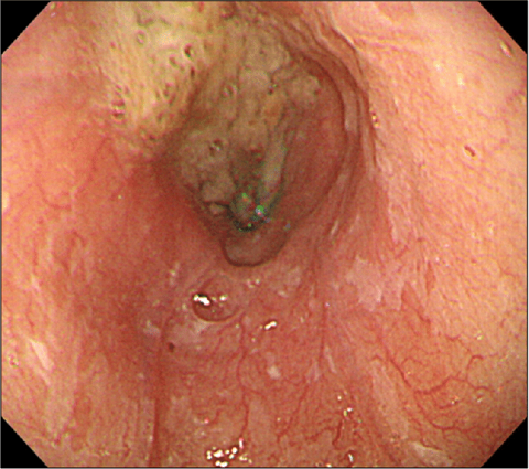
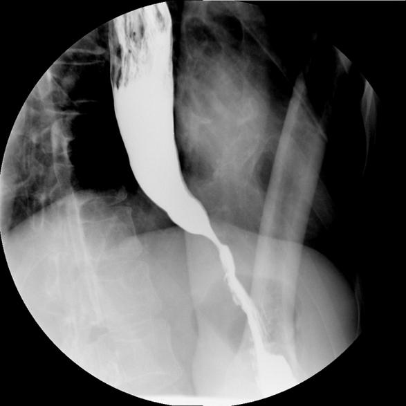
Before Treatment
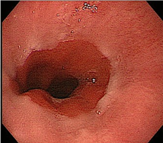
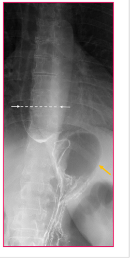
After Treatment
What Is Achalasia?
Achalasia is a rare swallowing disorder that affects the esophagus (the tube connecting the throat to the stomach).
In achalasia:
The esophagus muscles don’t work properly, so food doesn’t move down smoothly.
The lower esophageal sphincter (LES) (a ring of muscle at the bottom of the esophagus) stays too tight and doesn’t relax to let food enter the stomach.
Most common in adults between ages 30–60, especially in their 40s.
Occurs about twice as often in men than women.
What Causes It?
The exact cause is not known, but possible factors include:
Nerve cell damage in the esophagus.
Infections: Some patients show evidence of past viral or parasitic infections (e.g., measles, herpes, HPV).
Autoimmune disease: The body’s immune system may attack its own nerve cells.
People with achalasia are more likely to have conditions like type 1 diabetes, lupus, rheumatoid arthritis, or Sjögren’s syndrome.
Symptoms
Symptoms usually occur during or after eating and may include:
Trouble swallowing (food or liquid feels stuck in the chest) → most common symptom
Regurgitation: food or liquid coming back up into the mouth
Chest pain, sometimes severe enough to wake you at night
Heartburn
Coughing at night or choking
Aspiration (food or liquid entering the lungs)
⚠️ Severe achalasia can cause:
Weight loss and malnutrition
Fatigue
Pneumonia (if food enters the airways)
How Is It Different From GERD?
GERD (acid reflux): LES is too loose, letting stomach acid flow back up.
Achalasia: LES is too tight, preventing food from moving down.
Types of Achalasia
Doctors classify achalasia into three main types:
Type I (Classic achalasia)
Very little muscle movement in the esophagus.
Food moves mostly by gravity.
Type II
Pressure builds up in the esophagus, squeezing it.
Most common type and often causes more severe symptoms.
Type III (Spastic achalasia)
Abnormal, spasm-like contractions near the bottom of the esophagus.
Causes severe chest pain, sometimes mimicking a heart attack.
Considered the most severe form.
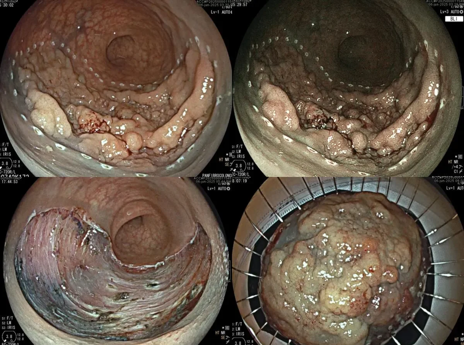
What Is Stomach Cancer?
Stomach cancer begins in the stomach.
The most common type is adenocarcinoma, which starts in the inner lining.
Other types (from the middle or outer stomach layers) are rare.
Although rates have gone down in recent decades, it is still a common cancer worldwide.
What Causes It?
The exact cause is not fully known, but certain factors increase the risk:
Diet: Eating many smoked, salted, or processed foods and too few vegetables.
Lifestyle: Smoking and drinking alcohol.
Infection: Helicobacter pylori (a bacteria that damages the stomach lining).
Other conditions: Stomach ulcers or abnormal cell growth.
Family history: Some inherited conditions.
⚠️ Stomach cancer can still occur even without any known risk factors.
Symptoms
Early stomach cancer may not cause symptoms. Some mild symptoms (such as fullness, stomach discomfort, or fatigue) can be mistaken for other conditions.
More concerning symptoms include:
Unexplained weight loss
Severe belly pain
Nausea and vomiting
Loss of appetite
Vomiting blood
Dark, sticky stools (tarry stool)
Trouble or painful swallowing
👉 If these symptoms last for more than two weeks, see a gastroenterologist.
How Is It Diagnosed?
At your appointment, the doctor will:
Ask about your health and symptoms.
Perform a physical exam.
Request blood and stool tests.
Additional tests may include:
CT (CAT) scan
PET scan
MRI
Upper endoscopy (EGD) → a camera examines the stomach.
Biopsy → a tissue sample taken during endoscopy.
Endoscopic ultrasound → checks how deep the cancer has grown.
What Is a Carcinoid Tumor?
Carcinoid tumors are rare, usually slow-growing tumors.
They can occur in different parts of the body and are considered cancerous, but many take years to develop.
Most people may not know they have one, but some tumors grow faster.
Rarely, if the tumor spreads, it may cause carcinoid syndrome, due to excess hormone release.
Where Carcinoid Tumors Can Occur
Common locations include:
Small intestine
Appendix
Rectum
Lungs
Colon
Stomach
Pancreas
Liver
Types of Carcinoid Tumors
Slow-growing tumors
Most common type, usually small (<1 cm)
Rarely spread
Faster-growing tumors
Can grow more quickly and spread to other areas
Hormone-secreting (functional) tumors
Produce hormones such as insulin, gastrin, serotonin
Can cause carcinoid syndrome
Non-hormone-secreting (nonfunctional) tumors
Most common
Do not produce hormones
Symptoms
Symptoms depend on location and hormone activity. Many tumors are nonfunctional and cause no symptoms.
Symptoms related to tumor size/location:
Abdominal pain or pressure
Changes in bowel habits
Blood in stool
Nausea or vomiting
Chest pain
Cough or blood-tinged sputum
Weight loss
Symptoms from hormone release:
Facial flushing
Diarrhea
High or low blood sugar
Ulcers or rashes
Anxiety, confusion, heart palpitations, fainting
Symptoms of carcinoid syndrome:
Facial flushing
Sweating
Wheezing or shortness of breath
Diarrhea
Heart palpitations or heart murmur
Low blood pressure
Confusion
Diagnosis
Carcinoid tumors are often found by accident during imaging for another reason. Diagnosis may include:
X-ray → basic imaging
CT scan → detailed cross-section images
MRI → detailed images using magnetic fields
DOTATATE PET/CT scan → highlights tumors using a special tracer
Biopsy → tissue sample to confirm diagnosis
Urine test → checks for hormone byproducts (5-HIAA)
Blood test → measures hormone levels or chromogranin A
Treatment
Small tumors that haven’t spread:
Surgery → often curative
Tumors that have spread or are too large:
Partial/debulking surgery → remove most tumor to control symptoms
Chemotherapy → drugs to target tumor cells
Octreotide → injection that reduces hormone production and slows growth
Liver-directed treatments → chemotherapy or blood-flow-blocking pellets if tumor spreads to liver
What Is Zenker’s Diverticulum?
Zenker’s Diverticulum is a small pouch that forms in the upper part of the esophagus (the tube connecting the throat to the stomach).
It occurs when the lining of the esophagus bulges outward through a weak spot in the muscle.
This condition can cause swallowing problems and other symptoms, especially in older adults.
Causes
Exact cause is not fully known.
Often linked to pressure changes in the throat or esophagus during swallowing.
Weakness in the esophageal muscles may allow a pouch to form over time.
Symptoms
Symptoms are usually related to food or liquid getting trapped in the pouch:
Trouble swallowing (dysphagia)
Feeling of food sticking in the throat
Regurgitation of undigested food, sometimes hours after eating
Bad breath (halitosis)
Coughing, choking, or aspiration (food/liquid entering the lungs)
Hoarseness or sore throat
Rarely, weight loss if swallowing becomes difficult over time
Complications
If untreated, Zenker’s diverticulum can cause:
Repeated lung infections (aspiration pneumonia)
Chronic bad breath
Malnutrition due to difficulty eating
Diagnosis
Diagnosis may include:
Barium swallow X-ray → patient swallows a contrast liquid to highlight the pouch on X-ray
Endoscopy → a small camera is used to look inside the esophagus
Esophageal manometry → measures pressure in the esophagus and throat muscles
Treatment
Treatment depends on symptoms and size of the pouch:
Mild cases:
Monitoring if symptoms are minimal
More severe cases:
Surgery → remove or reduce the pouch
Endoscopic procedures → less invasive options to divide the pouch from the esophagus, allowing better swallowing
Lifestyle adjustments → eating slowly, chewing food thoroughly, and drinking plenty of liquids with meals
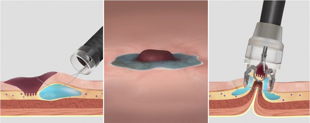
POEM Procedures:
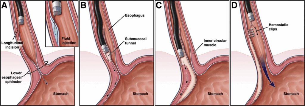
Advanced Procedures:
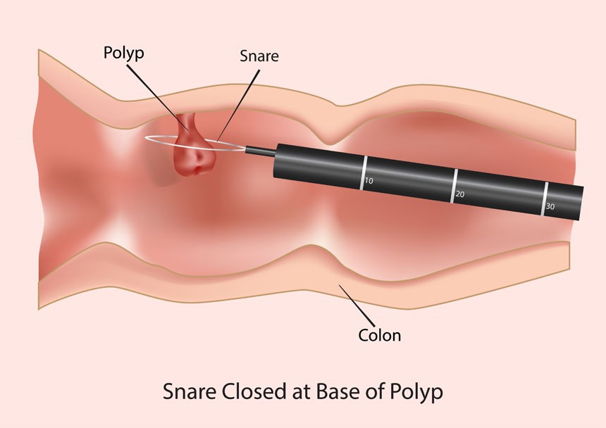
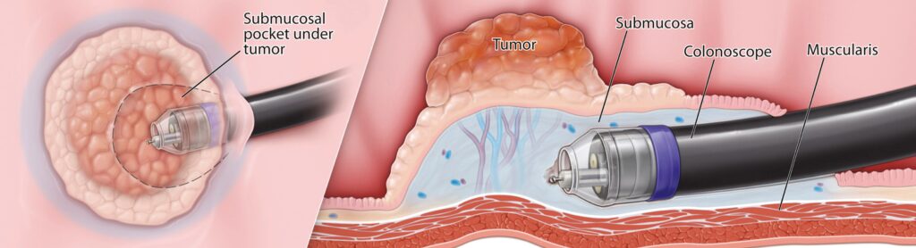
IBD and Immunology Clinic
Key Conditions Treated:
Read MoreLess
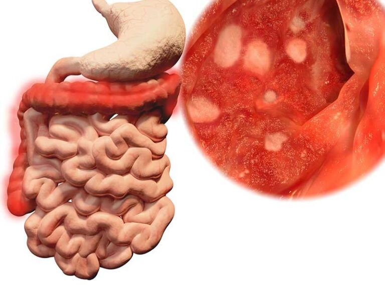
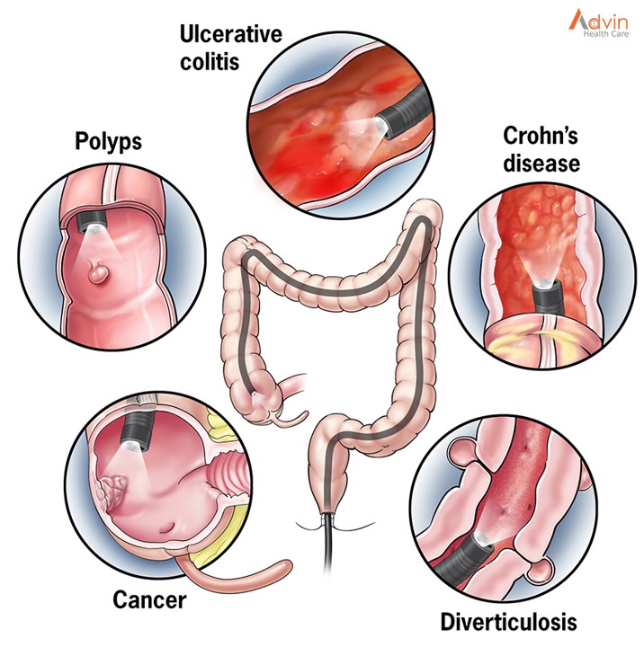
What Is Crohn’s Disease?
Crohn’s disease is a chronic inflammatory disease of the gastrointestinal (GI) tract.
It is an autoimmune disorder, meaning the immune system mistakenly attacks healthy tissue.
The disease can come and go, affecting different parts of the GI tract over time.
Who Gets Crohn’s Disease?
Often begins in teens or twenties, but can appear earlier or later.
About 1 in 6 patients show symptoms before age 15.
Genetics may play a role — family members may also be affected.
More common in the Jewish population than the general population.
Symptoms
Crohn’s disease affects people differently, depending on the part of the GI tract involved. Common symptoms include:
Abdominal cramps
Diarrhea
Weight loss
Fever
Anemia
Delayed growth in children (may appear before GI symptoms)
Non-intestinal symptoms
Some patients experience problems outside the gut, including:
Colitic arthritis → joint pain in knees, ankles, hips, wrists, and elbows
Pericholangitis → inflammation around bile ducts
Kidney stones
Urinary tract complications
Fistulas → abnormal connections between organs or skin
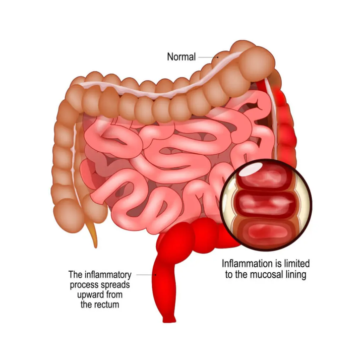
What Is Ulcerative Colitis?
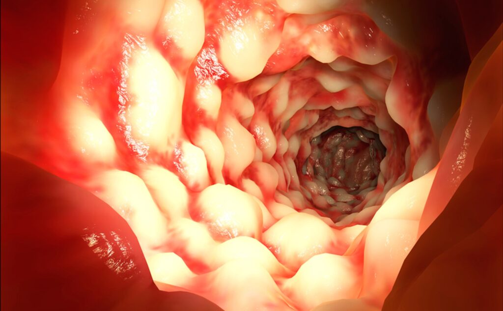
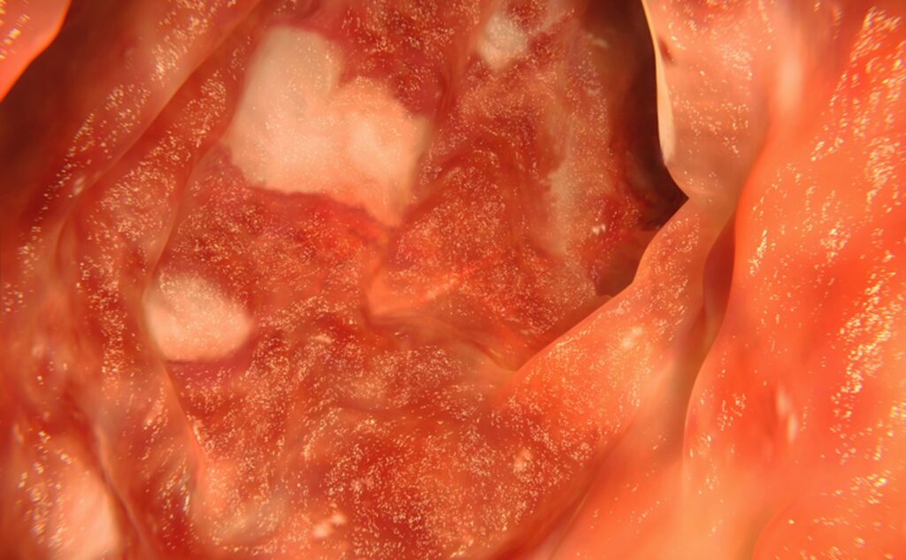
Ulcerative colitis is an inflammatory bowel disease (IBD).
It causes inflammation of the inner lining of the large intestine (colon) and rectum.
The severity and extent of inflammation can vary from person to person.
Difference from Crohn’s disease:
Ulcerative colitis affects only the inner lining of the bowel.
Crohn’s disease can affect all layers of the bowel wall.
Symptoms
Symptoms may include:
Bloody diarrhea (often the main symptom)
Frequent bowel movements
Abdominal or rectal pain
Fever
Weight loss
Joint pain
Skin rashes
Occasionally: constipation or rectal spasm
What Is Celiac Disease?
Celiac disease is a chronic digestive disorder that damages the small intestine.
It stops the body from absorbing nutrients from food properly.
It is not the same as gluten sensitivity or intolerance.
What Causes It?
Celiac disease is genetic — it can run in families.
It occurs in people who are allergic to gluten, a protein found in:
Wheat
Rye
Barley
Sometimes in small amounts in oats
When people with celiac disease eat gluten, the immune system attacks the small intestine, damaging tiny finger-like bumps called villi.
Villi absorb nutrients from food. Without them, your body can’t get enough nutrients, no matter how much you eat.
Symptoms may be triggered or worsened by:
Stress
Pregnancy or childbirth
Surgery or injury
Infection
Who Is More Likely to Have Celiac Disease?
People of European ancestry
White individuals
Those with type 1 diabetes
People with Down syndrome
People with other autoimmune diseases
Individuals who are infertile
People with irritable bowel syndrome (IBS) with diarrhea
Symptoms
Symptoms vary from person to person, and some people may have no symptoms at all.
Digestive Symptoms:
Chronic diarrhea or constipation
Abdominal pain or bloating
Gas or pale, foul-smelling stool
Weight loss
Other Physical Symptoms:
Fatigue due to anemia (low blood count)
Tingling or numbness in legs
Weak, brittle bones or fractures (osteoporosis)
Missed menstrual periods
Infertility
Dental enamel problems
Pain Symptoms:
Muscle cramps or bone pain
Joint pain
Itchy, painful skin rash
Children-Specific Symptoms:
Delayed growth or failure to thrive
⚠️ Even without symptoms, celiac disease can still cause complications over time.
Advanced Procedures:
Acidity Reflux & GI Motility Clinic
Key Conditions Treated:
Read MoreLess
What Is GERD?
GERD happens when stomach contents flow back into the esophagus (the tube connecting your throat to your stomach).
Occasional reflux is common, but it becomes GERD when symptoms are:
Frequent
Severe
Or cause damage to the esophagus, throat, or lungs.
Main Symptoms
Heartburn → a burning or fiery feeling in the chest
Regurgitation → sour or bitter liquid coming up into the throat or mouth
The combination of heartburn and regurgitation is the most common sign of GERD.
Other Symptoms
Non-burning chest pain (often in the middle of the chest, sometimes spreading to the back)
Difficulty swallowing (dysphagia)
Throat, voice, and lung-related symptoms:
Sore throat
Coughing
Increased saliva
Shortness of breath
What Is Chronic Cough?
Coughing is your body’s way of clearing the throat and airways of irritants.
Normally, it helps protect the lungs.
Sometimes, coughing can become chronic (long-lasting) even when nothing needs to be cleared.
A cough is considered chronic if it lasts 8 weeks or more.
Constant coughing can irritate the vocal cords, making them overly sensitive and causing the urge to cough more often.
Common Causes of Chronic Cough
Medicines (especially certain blood pressure medications)
Lung conditions like asthma or COPD (chronic obstructive pulmonary disease)
Allergies or sinus problems causing postnasal drip
Acid reflux (GERD or laryngopharyngeal reflux)
Swallowing difficulties that allow food or liquids to enter the airway
Diagnosis
Chronic cough can be linked to the lungs, sinuses, allergies, or reflux.
Your ENT (ear, nose, and throat doctor) may refer you to:
A pulmonologist (lung specialist)
A gastroenterologist (digestive specialist)
Tests may be done to find the exact cause.
Treatment
Treatment depends on the cause and may include:
Medicines to manage reflux, allergies, asthma, or other conditions
Lifestyle changes to reduce irritation
Speech therapy with a speech-language pathologist to:
Improve vocal hygiene (reduce irritation of the voice box)
Learn to control cough triggers
Practice breathing and voice exercises to reduce coughing
Before Treatment
After Treatment
What Is Achalasia?
Achalasia is a rare swallowing disorder that affects the esophagus (the tube that carries food from the throat to the stomach).
In achalasia, the muscles of the esophagus don’t move food down properly.
The valve at the bottom of the esophagus (called the lower esophageal sphincter, or LES) doesn’t relax to let food enter the stomach.
This makes swallowing difficult and can cause food or liquids to come back up.
Achalasia usually affects adults between 30–60 years old and is about twice as common in men.
Possible Causes
The exact cause is unknown, but doctors believe achalasia may be linked to:
Nerve damage in the esophagus
Infections (such as viruses or parasites)
Autoimmune conditions, where the body attacks its own tissues
People with achalasia may also be at a higher risk of other autoimmune disorders (such as rheumatoid arthritis, lupus, or type 1 diabetes).
Symptoms of Achalasia
Symptoms often occur during or after eating. They may include:
Trouble swallowing (food or drink feels “stuck”) — most common symptom
Regurgitation (food or liquid coming back up into the mouth)
Chest pain (sometimes severe, may feel like a heart attack)
Heartburn
Coughing, especially at night
Choking or inhaling food/liquid into the airway (aspiration)
If achalasia becomes severe, it can lead to:
Weight loss
Fatigue
Malnutrition
Pneumonia (if food enters the lungs while coughing or choking)
Types of Achalasia
Doctors describe achalasia in three main types:
Type 1 (Classic achalasia): Esophagus muscles barely move; food goes down mostly by gravity.
Type 2: Pressure builds up in the esophagus, causing compression. This is the most common type.
Type 3 (Spastic achalasia): Muscles at the lower end of the esophagus contract abnormally, causing severe chest pain that can mimic a heart attack.
What Is Rumination Syndrome?
Rumination syndrome is a rare condition that affects both children and adults.
After eating, food comes back up into the mouth within 10–15 minutes.
This can continue for 1–2 hours after meals.
The food is undigested and often tastes the same as when it was first eaten.
A person may rechew and swallow the food or spit it out.
⚠️ Rumination is an automatic reflex, not something done on purpose.
What Causes It?
The exact cause isn’t fully known. Experts believe rumination is related to:
A learned reflex where the diaphragm (a muscle under the lungs) relaxes, pushing food back up.
It is similar to burping, but instead of gas, food comes up.
Rumination is unconscious — people don’t have control over it.
Symptoms of Rumination Syndrome
Food coming back up shortly after meals
Food tastes normal (not sour or bitter) because it hasn’t been digested
May happen at every meal, day after day
👉 Rumination is different from vomiting:
Vomiting brings up digested food or stomach acid (tastes bad).
Rumination brings up undigested food that tastes the same as when eaten.
What Is Bloating?
Bloating is when your belly feels full, tight, or swollen, often because of gas in your digestive system.
👉 It’s different from a belly that looks bigger due to loose abdominal muscles (common after pregnancy or with aging). Knowing the difference helps you get the right treatment.
Causes of Bloating
1. Constipation
One of the most common causes. Even if you have bowel movements, you may still be constipated. Signs include:
Straining during bowel movements
Hard, pebble-like stools
Feeling like you didn’t completely empty
When stool stays in the colon too long, bacteria break it down and create gas → bloating.
2. Gut Sensitivity (IBS)
People with Irritable Bowel Syndrome (IBS) may feel pain, cramping, diarrhea, and bloating even with small amounts of gas.
3. Small Intestinal Bacterial Overgrowth (SIBO)
Normally, few bacteria live in the small intestine.
With SIBO, extra bacteria grow there → gas and bloating.
More common in people with intestinal surgery or IBS with diarrhea.
4. Gastroparesis
A condition where the stomach empties too slowly.
Can cause bloating, nausea, and sometimes bowel blockages.
5. Gynecological Causes
Conditions involving the ovaries or uterus may cause bloating.
Regular pelvic exams are important to rule these out.
Advanced Procedures:
Emergency 24x7 Endoscopy Unit
Key Conditions Treated:
Read MoreLess
What Is a Mallory-Weiss Tear?
A Mallory-Weiss tear is a small cut in the tissue at the lower end of your esophagus (the tube that carries food from your throat to your stomach).
It usually happens after forceful vomiting or coughing.
The tear can cause bleeding inside your digestive tract.
Causes
The most common cause is violent coughing or vomiting.
Other, less common causes include:
Hiatal hernia (part of the stomach pushes into the chest)
Childbirth (from increased abdominal pressure)
Symptoms
A Mallory-Weiss tear may not always be obvious, but possible signs include:
Vomit that is bright red or looks like coffee grounds
Black, tar-like stools
Blood in stools
Weakness, dizziness, or fainting
Shortness of breath
Pale skin
Abdominal or chest pain
Diagnosis
Your doctor may:
Test your stool for blood
Do an endoscopy: a flexible tube with a camera is placed through your mouth into your esophagus to look for the tear
Treatment
In most cases, the tear heals on its own without treatment.
If bleeding continues, treatment may include:
Endoscopic injection (to stop bleeding)
Heat treatment (to seal the blood vessel)
Clips placed with the endoscope to close the tear
What Are Black Stools?
Black stools are bowel movements that look very dark, tar-like, and sticky. The medical term for this is melena.
They are usually caused by bleeding in the upper digestive tract, such as the stomach or esophagus.
Sometimes, however, black stools can happen from harmless causes, such as food or medicine.
Common Causes
Serious Causes (need medical attention)
Stomach ulcers
Mallory-Weiss tear (a tear in the esophagus)
Gastritis (stomach inflammation)
Esophageal or stomach cancer
Bleeding blood vessels in the stomach or esophagus
Harmless Causes
Iron supplements
Certain foods (black licorice, blueberries, dark chocolate)
Medicines like Pepto-Bismol® (bismuth subsalicylate)
Symptoms
If your black stool is due to bleeding, you may also notice:
Dizziness or fainting
Weakness
Shortness of breath
Abdominal pain
Vomiting blood (bright red or coffee-ground–like)
Pale skin
What Is Foreign Body Ingestion?
Foreign body ingestion happens when food, toys, coins, or other small objects get stuck in a child’s airway or digestive tract (throat, esophagus, windpipe, or lungs).
Children under 3 years old are especially at risk because they explore by putting things in their mouths.
Prevention Tips
Keep small toys and objects out of reach (always check warning labels).
Do not give nuts, sunflower seeds, raw carrots, or popcorn kernels to young children.
Always supervise children while eating or playing.
Teach older children not to put small items in their mouths.
🚨 Prevention is the best way to avoid accidents!
Symptoms
A child may have swallowed or inhaled something if you notice:
Trouble breathing or speaking
Drooling or vomiting
Chest or stomach pain
Persistent cough or wheezing (especially on one side)
Bluish skin (cyanosis) from lack of oxygen
Pneumonia that does not improve
If the object is in the nose, symptoms may include:
Foul-smelling mucus
Bloody discharge from one nostril
Difficulty breathing through that side of the nose
Advanced Procedures:
GI Functional & Physiology Testing Clinic
Key Conditions Treated:
Read MoreLess
What is IBS?
Irritable bowel syndrome (IBS) is a common condition that affects the large intestine (colon).
It causes abdominal pain or discomfort and changes in bowel habits (diarrhea, constipation, or both).
IBS is different from inflammatory bowel disease (IBD), which causes lasting inflammation in the intestines.
Causes
IBS is related to oversensitive nerves in the gut, which may cause the intestines to react strongly to normal processes.
In some people, IBS may be linked to how the gut and brain communicate.
It can sometimes develop after a severe intestinal infection (post-infectious IBS).
Family history may increase risk.
Who Gets IBS?
IBS affects up to 15% of people in the U.S.
Women are about twice as likely to have IBS as men.
Symptoms usually start before age 40, sometimes in childhood or young adulthood.
Types of IBS
There are four main types:
IBS-D: Mostly diarrhea with abdominal discomfort
IBS-C: Mostly constipation with abdominal discomfort
IBS-M (mixed): Alternating diarrhea and constipation
IBS-U: Symptoms vary and do not fit the other types
Common Symptoms
Digestive Symptoms:
Abdominal pain or cramping
Bloating, fullness, or burning
Mucus in the stool
Feeling of urgency or incomplete bowel movements
Other Symptoms:
Some people also experience:
Migraines
Sleep disturbances
Anxiety or depression
Chronic pelvic pain
Fibromyalgia
Symptoms may come and go, and stress, certain foods, or meals can sometimes trigger them.
How IBS Affects Daily Life
Some people can manage symptoms and continue their normal activities.
Others may find that IBS limits daily life, including work and social activities.
Symptoms may improve when stress is reduced or may appear randomly.
What is Constipation?
Constipation happens when:
Bowel movements are infrequent (less than 3 times a week)
Stool is hard, dry, or in small pieces
You strain or feel pain while passing stool
Normal bowel habits vary. Some people go 3 times a day, while others go 3 times a week.
What Causes Constipation?
Constipation occurs when the colon absorbs too much water, making stool hard and difficult to pass.
Common diet and lifestyle causes include:
Not enough exercise
Not drinking enough fluids
Not eating enough fiber-rich foods
Ignoring the urge to have a bowel movement
Changes in routine (travel, pregnancy, aging)
Other possible causes:
Certain medications
Irritable bowel syndrome (IBS) or other bowel problems
Slow intestinal movement
Metabolic issues, like thyroid problems
Intestinal blockage
Symptoms of Constipation
Fewer than 3 bowel movements per week
Hard, dry, or lumpy stool
Straining or pain when passing stool
Feeling bloated or full
Belly discomfort or cramps
What is SIBO?
SIBO happens when too many bacteria grow in the small intestine, which normally has fewer bacteria than the large intestine. This can interfere with digestion and nutrient absorption.
Causes of SIBO
SIBO can occur when the small intestine doesn’t move food and bacteria normally, leading to bacterial overgrowth. Common causes include:
Previous intestinal surgery
Irritable bowel syndrome (IBS) or other gut disorders
Slow intestinal movement
Structural problems in the intestines
Symptoms of SIBO
Symptoms may vary but often include:
Bloating and feeling full
Gas and belching
Diarrhea or constipation
Abdominal pain or cramps
Nausea
Unintended weight loss (in severe cases)
SIBO symptoms can be similar to other digestive conditions, so proper diagnosis is important.
How is SIBO Diagnosed?
Doctors may use:
Breath tests to detect bacterial activity
Blood tests to check for nutrient deficiencies
Stool tests in some cases
Imaging or endoscopy if needed to look for underlying causes
Treatment of SIBO
Treatment usually targets the bacterial overgrowth and the underlying cause:
Antibiotics to reduce bacteria
Dietary changes, such as low FODMAP diet
Probiotics in some cases
Treating underlying disorders, like IBS or motility issues
What is Fecal Incontinence?
Fecal incontinence is the inability to control bowel movements, causing stool to leak unexpectedly from the rectum. It can range from occasional minor leaks to complete loss of control.
Causes of Fecal Incontinence
Fecal incontinence can happen due to problems with the muscles or nerves that control bowel movements. Common causes include:
Weak pelvic floor muscles
Damage to the anal sphincter from childbirth, surgery, or injury
Nerve damage from diabetes, stroke, or spinal cord injury
Constipation causing stool to harden and stretch the rectum
Diarrhea or loose stool that is hard to control
Aging, which can weaken muscles and nerves
Symptoms of Fecal Incontinence
Symptoms may include:
Accidental leakage of stool
Urgent need to go without enough time to reach the bathroom
Difficulty controlling gas
Skin irritation around the anus from frequent leakage
Severity varies—some people have occasional leakage, while others may experience frequent accidents.
How is Fecal Incontinence Diagnosed?
Doctors will usually:
Take a medical history and ask about bowel habits
Perform a physical exam, including checking anal muscles and nerves
Order tests such as anorectal manometry, imaging, or colonoscopy if needed
Treatment of Fecal Incontinence
Treatment depends on the cause and severity:
Dietary changes (more fiber or managing diarrhea)
Pelvic floor exercises (Kegel exercises)
Medications for constipation or diarrhea
Biofeedback therapy to strengthen muscles
Surgery in severe cases to repair muscles or nerves
What is Anorectal Dysfunction?
Anorectal dysfunction refers to problems with the muscles or nerves in the rectum and anus, which can affect bowel movements, stool control, and comfort during defecation.
Causes of Anorectal Dysfunction
Problems in this area can result from:
Muscle weakness in the pelvic floor or anal sphincter
Nerve damage from surgery, injury, or neurological conditions
Chronic constipation or straining
Aging, which can weaken muscles and reduce nerve function
Pelvic floor disorders
Symptoms of Anorectal Dysfunction
Symptoms may include:
Difficulty passing stool (constipation)
Straining or feeling of incomplete emptying
Stool leakage or fecal incontinence
Pain or discomfort in the rectum
Urgency or inability to control bowel movements
Rectal prolapse or bulging
Symptoms vary depending on the underlying cause and severity.
How is Anorectal Dysfunction Diagnosed?
Your healthcare provider may perform:
Medical history review and symptom discussion
Physical exam, including the pelvic floor and anal muscles
Anorectal manometry to measure muscle function
Defecography (imaging while passing stool)
Colonoscopy or imaging to rule out other conditions
Treatment of Anorectal Dysfunction
Treatment depends on the cause and severity:
Dietary changes (high-fiber diet, hydration)
Pelvic floor physical therapy and exercises
Biofeedback therapy to improve muscle coordination
Medications to manage constipation or diarrhea
Surgery for structural problems, severe prolapse, or sphincter repair
Advanced Procedures:
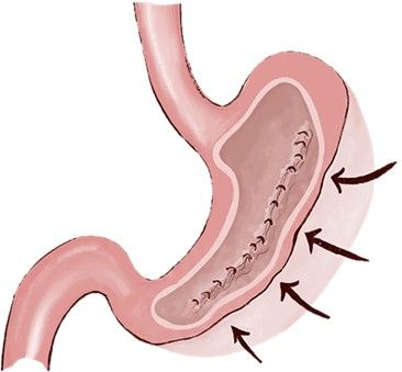
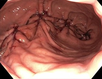
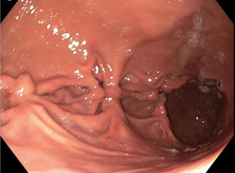
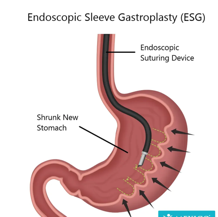
Bariatric and Metabolic Endoscopy
Advanced Procedures:
Capsule Endoscopy & Small Bowel Clinic
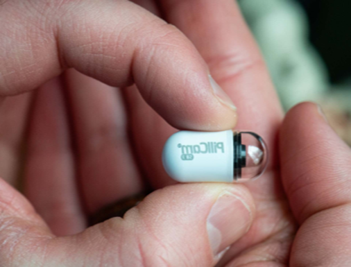
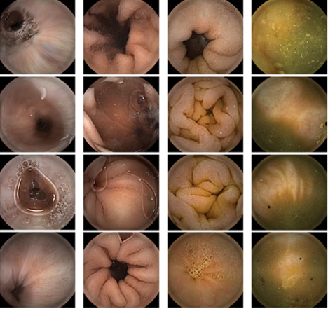
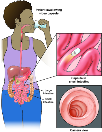
Key Conditions Treated:
Read MoreLess
What is GI Bleeding?
Gastrointestinal (GI) bleeding means bleeding anywhere in the digestive tract, which includes the esophagus, stomach, small intestine, large intestine (colon), rectum, and anus.
If the bleeding is obvious, you may see red or black blood in your stool or vomit.
If the bleeding is hidden (occult bleeding), you may not notice it at all. Special stool tests can detect this type.
Sometimes, the cause of bleeding is not immediately found — this is called obscure GI bleeding.
Symptoms of GI Bleeding
Bleeding may look different depending on where it comes from:
Bright red blood on or mixed with stool
Dark red or maroon-colored stool
Black, tar-like stool (called melena)
Bright red blood in vomit
Vomit that looks like coffee grounds
Other possible signs:
Change in bowel habits or stool appearance
Abdominal pain or tenderness
Weakness, dizziness, or fatigue from blood loss
Causes of GI Bleeding
Bleeding can be caused by many conditions, such as:
Stomach or small intestine ulcers
Inflammation of the stomach or colon
Small growths (polyps)
Diverticulosis (small pouches in the colon)
Blood vessel problems in the intestines
Tumors
Even foods and medicines can make stool look red or black (for example, beets, iron, or Pepto-Bismol), so your doctor will test your stool to confirm whether blood is present.
Why Diagnosis is Important
Finding the source of bleeding is important to prevent further blood loss and treat the underlying condition. Your doctor may recommend:
Blood tests
Stool tests for hidden blood
Endoscopy or colonoscopy (camera test)
Imaging studies if needed
What Is Crohn’s Disease?
Crohn’s disease is a chronic inflammatory disease of the gastrointestinal (GI) tract.
It is an autoimmune disorder, meaning the immune system mistakenly attacks healthy tissue.
The disease can come and go, affecting different parts of the GI tract over time.
Who Gets Crohn’s Disease?
Often begins in teens or twenties, but can appear earlier or later.
About 1 in 6 patients show symptoms before age 15.
Genetics may play a role — family members may also be affected.
More common in the Jewish population than the general population.
Symptoms
Crohn’s disease affects people differently, depending on the part of the GI tract involved. Common symptoms include:
Abdominal cramps
Diarrhea
Weight loss
Fever
Anemia
Delayed growth in children (may appear before GI symptoms)
Non-intestinal symptoms
Some patients experience problems outside the gut, including:
Colitic arthritis → joint pain in knees, ankles, hips, wrists, and elbows
Pericholangitis → inflammation around bile ducts
Kidney stones
Urinary tract complications
Fistulas → abnormal connections between organs or skin
What are Small Bowel Tumors?
The small bowel (small intestine) is the long tube that connects the stomach to the large intestine. It absorbs nutrients from food.
Small bowel tumors are growths that form in the lining or wall of the small intestine.
They can be benign (noncancerous) or malignant (cancerous).
Small bowel tumors are rare compared to tumors in the colon or stomach.
Types of Small Bowel Tumors
The most common types include:
Adenocarcinoma – starts from the lining of the intestine
Carcinoid (neuroendocrine) tumors – slow-growing tumors that may produce hormones
Lymphoma – cancer of the immune system that can affect the small bowel
Sarcoma/GIST (gastrointestinal stromal tumor) – tumors that develop in the wall of the small intestine
Symptoms
Small bowel tumors can be hard to detect early because symptoms are often vague. Possible symptoms include:
Abdominal pain or cramping
Nausea or vomiting
Unexplained weight loss
Blood in stool (red or black/tarry stool)
Anemia (low red blood cell count, causing fatigue or weakness)
Changes in bowel habits (diarrhea, constipation, or blockage)
Diagnosis
If your doctor suspects a small bowel tumor, they may order:
Blood tests (to check for anemia or other signs)
Endoscopy or capsule endoscopy (swallowing a small camera)
CT or MRI scans (detailed imaging of the abdomen)
Biopsy (taking a tissue sample to confirm diagnosis)
Treatment
Treatment depends on the type, size, and stage of the tumor:
Surgery – often the main treatment for removal
Chemotherapy or radiation – may be used for certain cancers like lymphoma
Targeted therapies or hormone treatments – for tumors like carcinoid or GIST
Regular follow-up is important to monitor for recurrence or new growths
What is Iron-Deficiency Anemia?
Iron-deficiency anemia is the most common type of anemia worldwide. It happens when your body does not have enough iron to make healthy red blood cells.
Red blood cells contain hemoglobin, which carries oxygen throughout the body.
Without enough iron, your body makes fewer and smaller red blood cells, leading to fatigue and other health problems.
What Causes Iron-Deficiency Anemia?
Iron deficiency can develop for several reasons:
Low iron in the diet – not eating enough iron-rich foods
Body changes – higher iron needs during growth spurts, pregnancy, or breastfeeding
Poor absorption – problems in the stomach or intestines, or surgeries that affect absorption
Blood loss – heavy periods, gastrointestinal bleeding, or injury
Symptoms
Iron-deficiency anemia can cause:
Pale skin
Fatigue or low energy
Fast heartbeat
Irritability
Sore or swollen tongue
Enlarged spleen
Cravings for non-food items like dirt or ice (pica)
⚠️ Symptoms can be similar to other conditions — always see your doctor for proper diagnosis.
How is it Diagnosed?
Doctors may use:
Blood tests – to check hemoglobin and iron levels
Stool or endoscopy tests – to look for hidden blood loss in the digestive tract
Rarely, bone marrow tests – to look at blood cell production
Treatment
Treatment depends on your age, health, and the cause of anemia. Options include:
1. Iron-Rich Diet
Eat more foods high in iron, such as:
Red meat, poultry, and liver
Fish and shellfish (clams, mussels, sardines)
Leafy greens (broccoli, kale, collards)
Beans, peas, and lentils
Iron-fortified cereals, bread, pasta, or rice
2. Iron Supplements
Usually taken by mouth for several months
Work best on an empty stomach or with orange juice (vitamin C helps absorption)
May cause dark stools or mild stomach upset
Some people may need IV iron if supplements don’t work
3. Treating the Underlying Cause
If anemia is due to blood loss, your doctor may recommend tests like an endoscopy or colonoscopy to find the source
Advanced Procedures:
Medical Directory Services
A Comprehensive Directory For Your Health Care.

Pancreatobiliary Clinic
Expert care for pancreatic, bile duct, and gallbladder disorders using advanced imaging and endoscopic techniques.

Flexible Endoscopic Surgery & Oncology
IBD and Immunology Clinic

Acidity Reflux & GI Motility Clinic

Emergency 24x7 Endoscopy Unit

GI Functional & Physiology Testing Clinic

Bariatric and Metabolic Endoscopy

Capsule Endoscopy & Small Bowel Clinic
We have 8+ more Care Service including Emergency Department. View All
why choose us
Experienced and Qualified Medical Professionals.
Eros lobortis imperdiet eu aliquet, aliquet fames. Molestie morbi lacinia mi magna lacinia odio. Sociosqu velit ullamcorper blandit aliquam dui commodo vivamus.
01
Trust
Multi-generational trust with over two decades of service
02
Advance Technology
Leading-edge technology and advanced endoscopic therapies
03
Holistic Approach
Holistic, patient-centered, ethical care approach
our services
Care For Popular Treatment.
Pancreatobiliary Clinic
Pancreatitis management
Stone removal (ERCP, EUS)
Bile duct stenting



GI Functional & Physiology Testing Clinic
IBS assessment
Manometry tests
Biofeedback therapy
Media & Public Appearances
Sharing Knowledge Beyond the Clinic


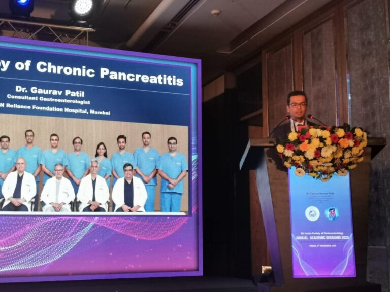
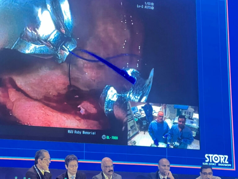
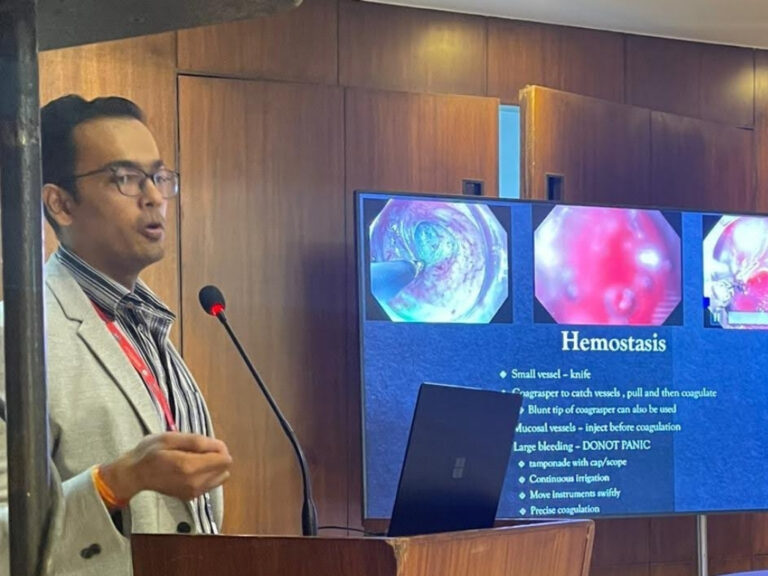



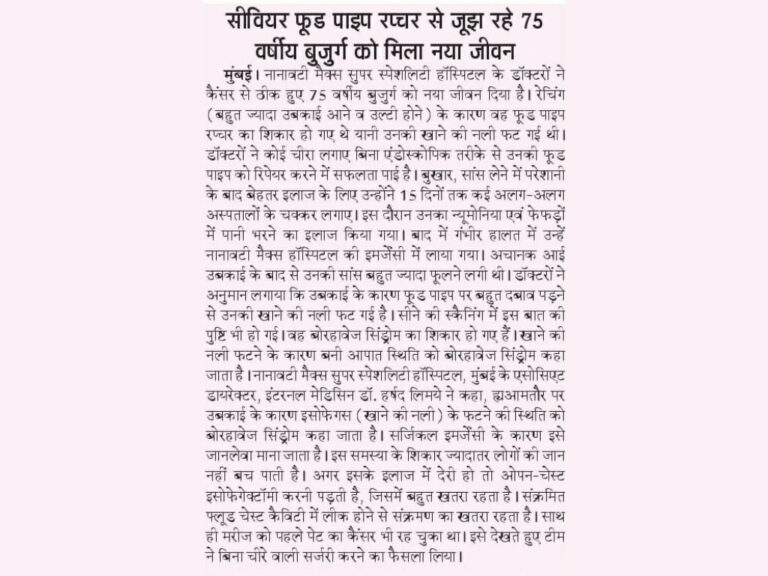
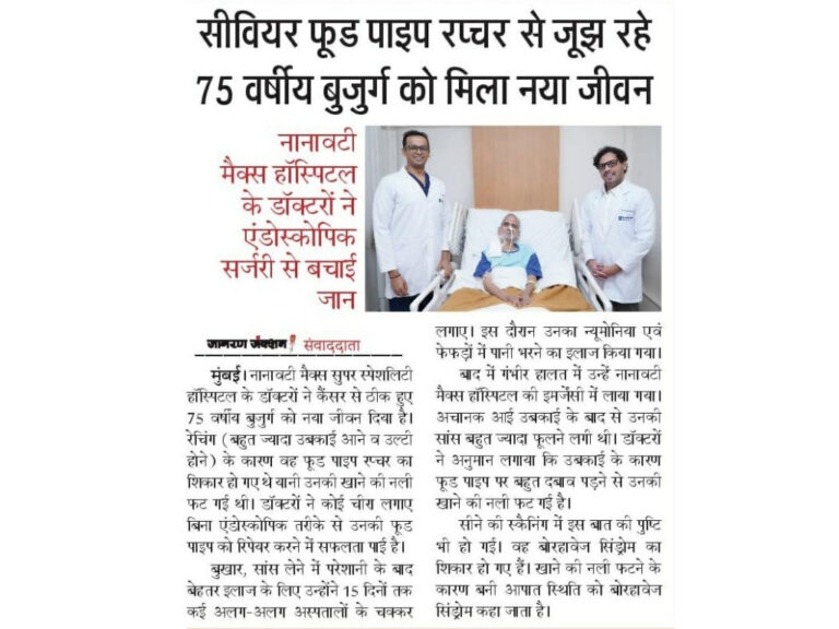
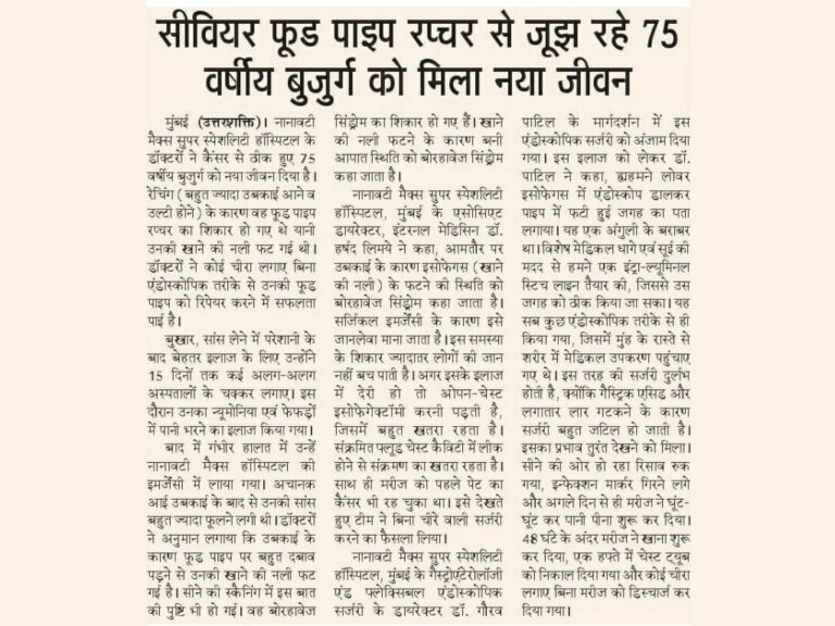
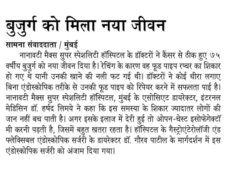
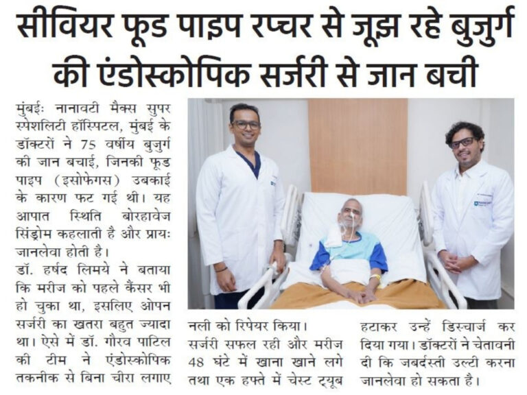
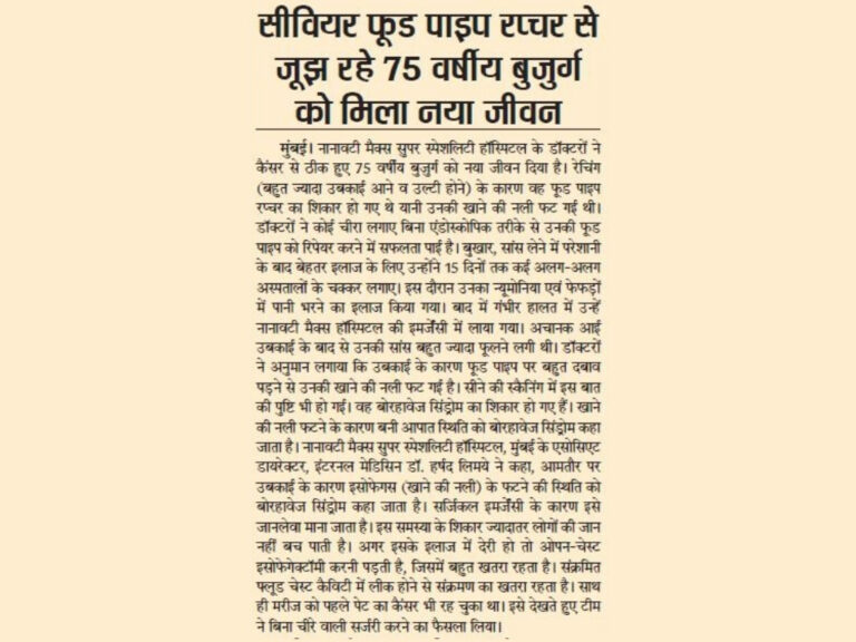
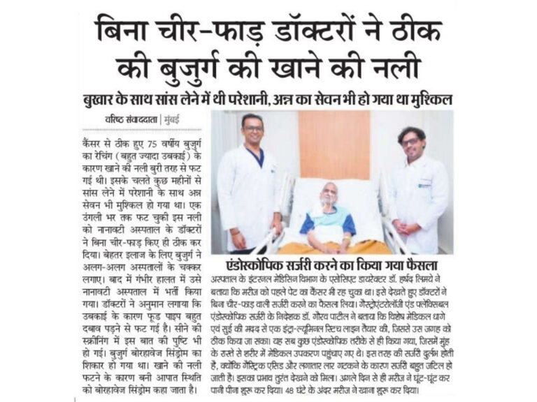
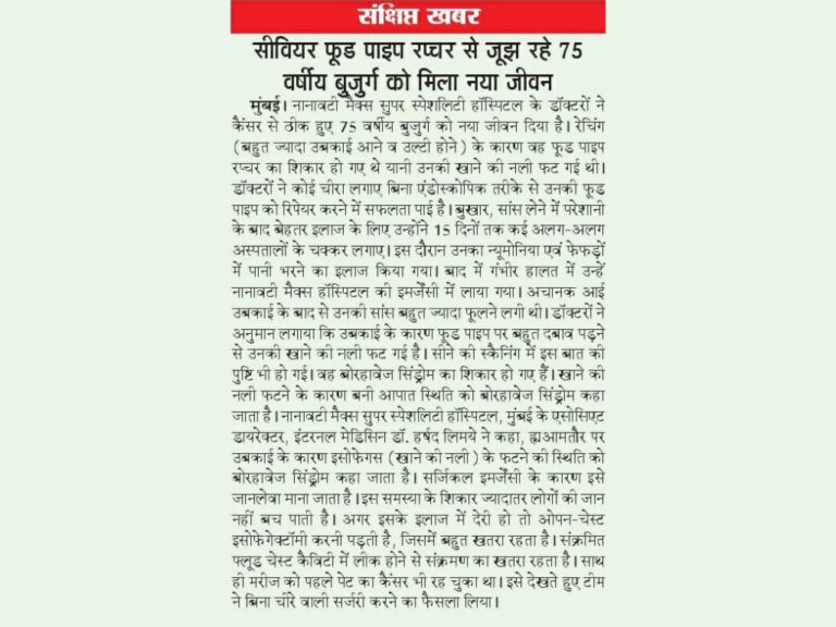
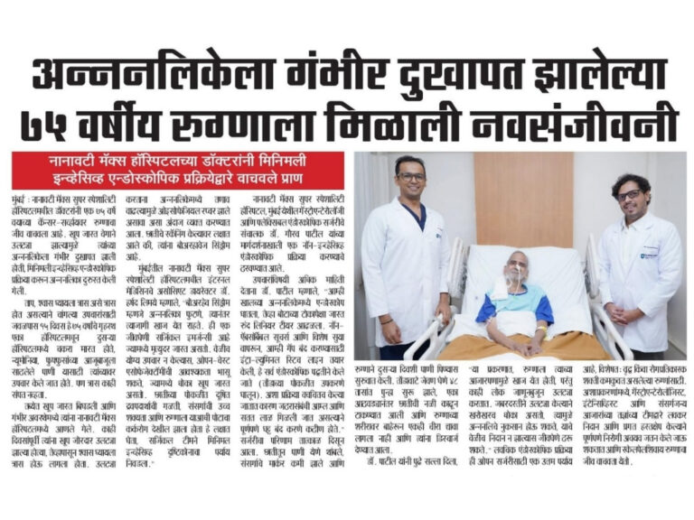
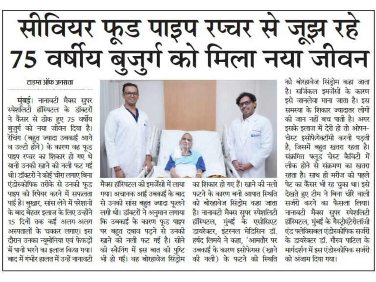




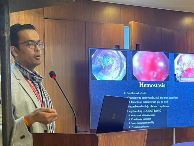
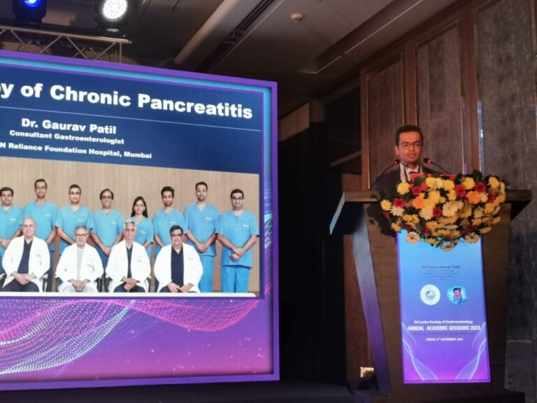


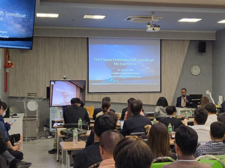

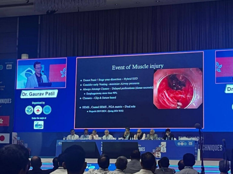

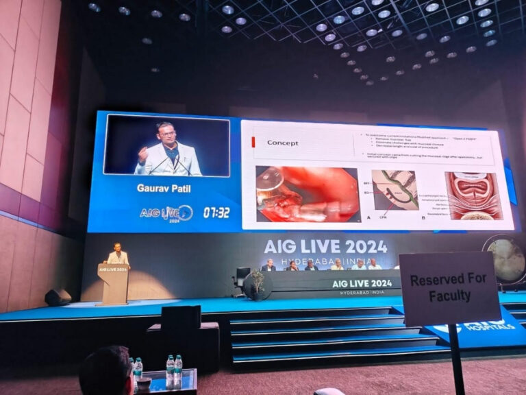

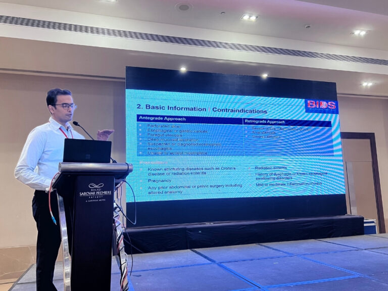

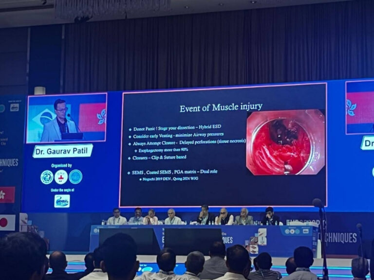



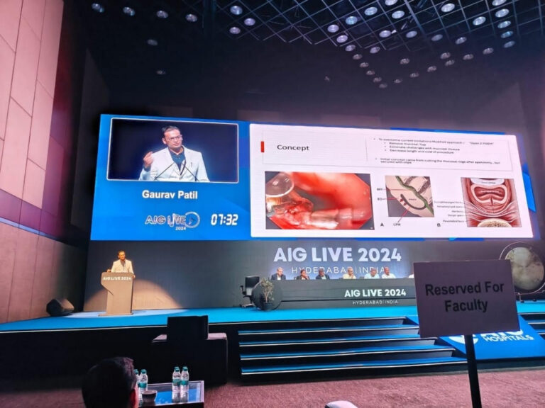


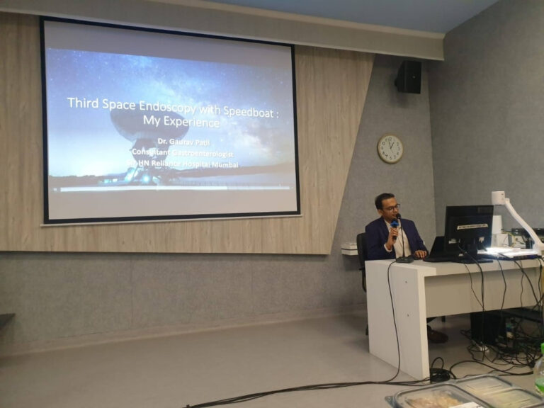
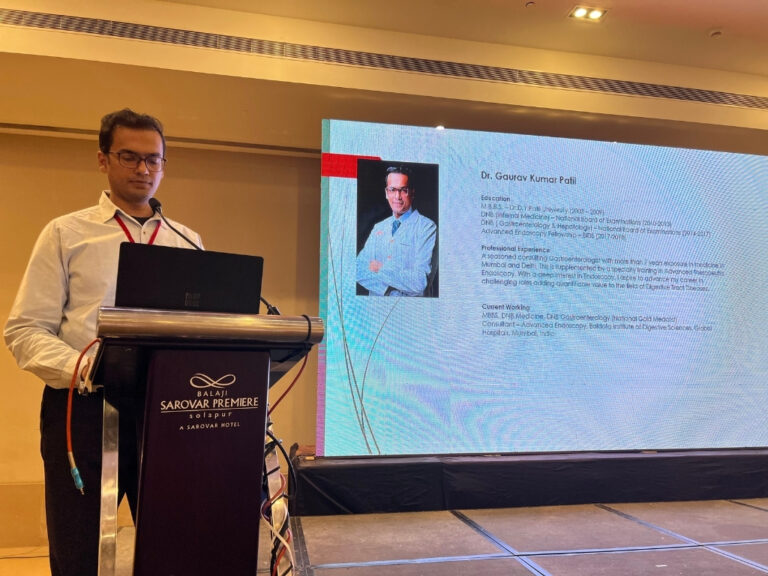








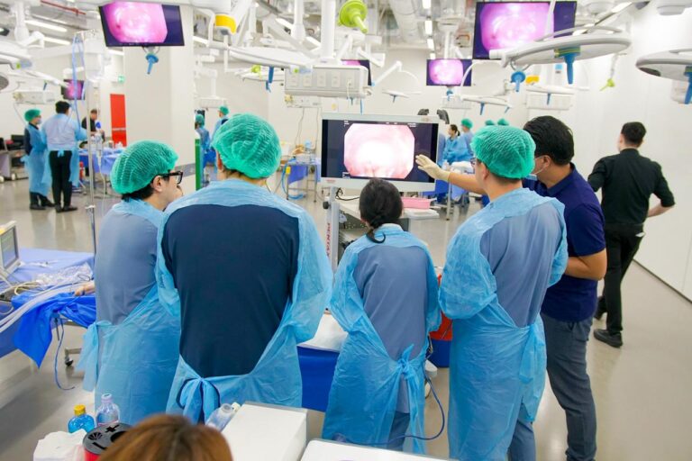
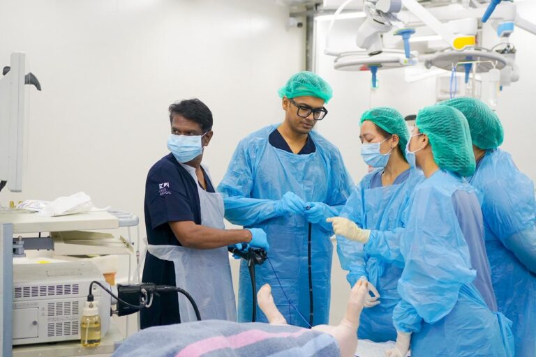




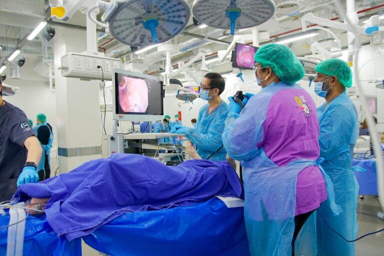

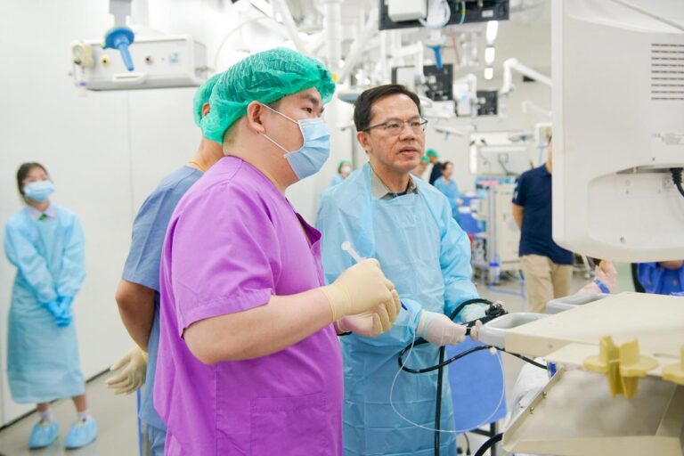


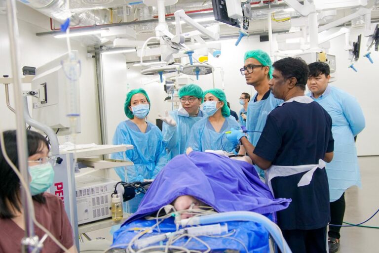

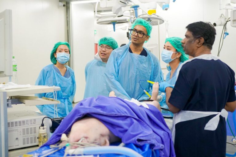
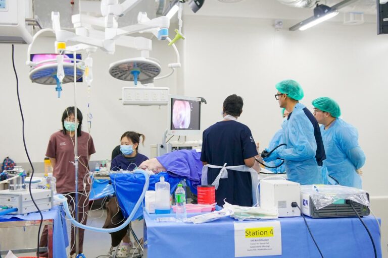
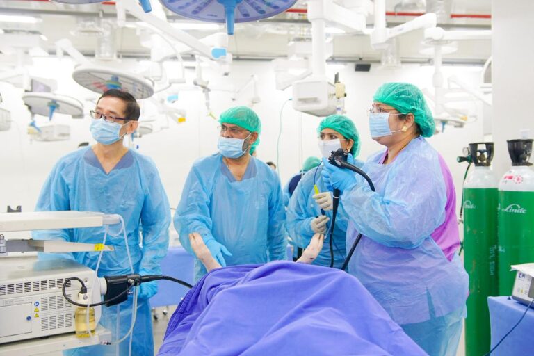

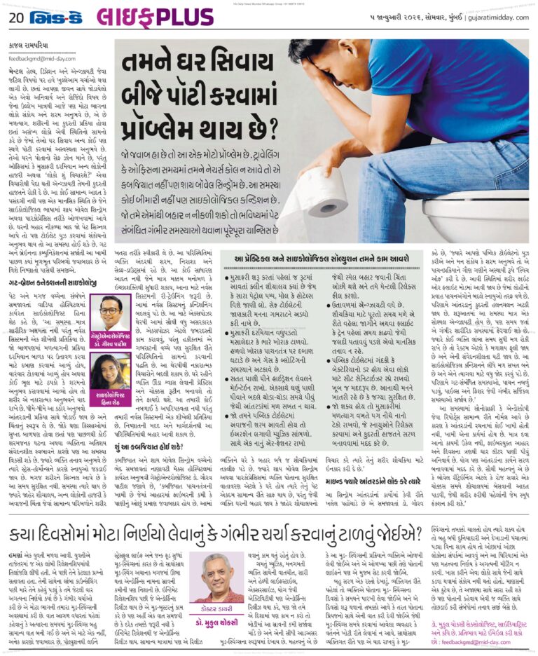
Expert guidance, tips, and updates for a healthier gut and better living
Healthy Gut Reads
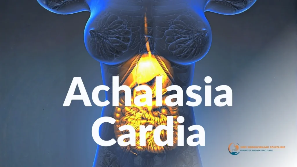
Achalasia Cardia: Complete Guide to Symptoms, Diagnosis & Advanced POEM Treatment in Malad, Mumbai Achalasia cardia is a rare condition that affects swallowing. It happens…
Advanced Role of EUS in Tumor Diagnosis & Precise Tissue Acquisition EUS for Diagnosis of Pancreatic and Biliary Tumors Endoscopic Ultrasound (EUS) plays a critical…
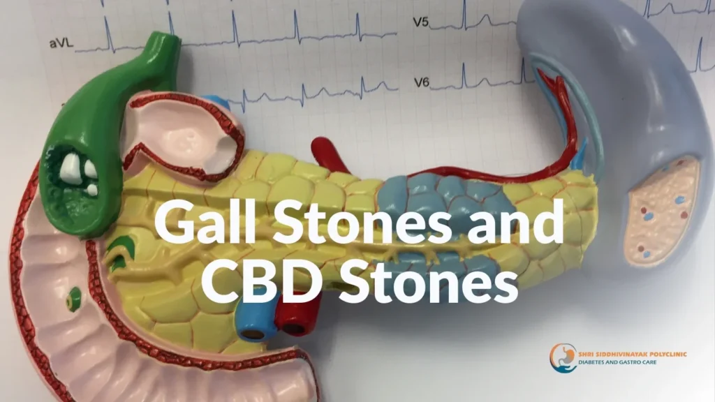
Gall Stones and CBD Stones: Simple Guide to Safe & Advanced Treatment in Malad West Gall stones and CBD stones are common digestive problems that…
Contact Us
Quick Contact
OUR LOCATION
Need a Doctor for Check-up? Call for an Emergency Service!
-
Pancreatitis Treatment in
Malad West
- Pancreatic Cancer Management by Dr. Gaurav Patil in Malad West
-
Gallstones Treatment in
Malad West -
Cholecystitis Care
Dr. Gaurav Patil - Endoscopic Retrograde Cholangiopancreatography
-
EUS (Endoscopic Ultrasound) by
Dr. Gaurav Patil - Achalasia Cardia Treatment in Malad West
- POEM (Peroral Endoscopic Myotomy) Dr. Gaurav Patil
-
Polyp Removal in
Malad West - Gastrointestinal Bleeding Management by Dr. Gaurav Patil
-
Varix / Varices Treatment in
Malad West -
Tumor Diagnosis & Treatment by
Dr. Gaurav Patil - Neuroendocrine Tumor (NET) Care Malad West
- GIST (Gastrointestinal Stromal Tumor) Treatment in Malad West
- Heartburn / Acidity Management by Dr. Gaurav Patil
-
Reflux / GERD Treatment in
Malad West
-
ARMS (Endoscopic Resection) by
Dr. Gaurav Patil - Bloating & Indigestion Management Malad West
- Obesity & Weight Loss Solutions in Malad West
-
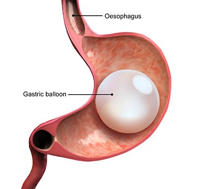
Intragastric Balloon Therapy by
Dr. Gaurav Patil - ESG (Endoscopic Sleeve Gastroplasty) Malad West
-
Bile Duct Stone Removal in
Malad West -
Cholangiocarcinoma Management
Dr. Gaurav Patil -
Laser Therapy in
Malad West
- ESWL (Extracorporeal Shock Wave Lithotripsy) by Dr. Gaurav Patil
- APC (Argon Plasma Coagulation) Malad West
-
Gastritis Treatment in
Malad West -
Ulcer Treatment & Care by
Dr. Gaurav Patil -
Gut Health in
Malad West -
Gut Wellness
Dr. Gaurav Patil -
Gut Microbiome
Malad West -
Brain Gut Axis Dr. Gaurav Patil
Malad West